Serviços Personalizados
Journal
artigo
Indicadores
Compartilhar
Journal of Human Growth and Development
versão impressa ISSN 0104-1282versão On-line ISSN 2175-3598
J. Hum. Growth Dev. vol.28 no.2 São Paulo maio/ago. 2018
https://doi.org/10.7322/jhgd.125609
ORIGINAL ARTICLE
DOI: http://dx.doi.org/10.7322/jhgd.125609
Clinical decision making for diagnosis and treatment of dental enamel injuries
Izabel Barzotto; Lilian Rigo
Faculdade Meridional (IMED) - Passo Fundo (RS), Brasil
ABSTRACT
INTRODUCTION: In general, there are difficulties in the decision making regarding the differential diagnosis and the most appropriate treatment in the lesions that affect the dental tissues by dentists, due to the fact that lesions in enamel have similar clinical characteristics
OBJECTIVE: To evaluate the correct decision making for the diagnosis and treatment of dental enamel lesions by professors and students of the Dentistry course
METHODS: Descriptive quantitative study, whose sample was composed by 98 students enrolled in the disciplines of Dental Clinics from IV to VIII level and by 23 professors. The instrument of data collection was a questionnaire composed of photographs of four clinical cases, whose teeth presented different lesions in dental enamel: dental enamel hypoplasia, dental fluorosis, amelogenesis imperfecta and dental caries
RESULTS: Of the 98 students, the predominant success was dental fluorosis, where 93.9% answered the diagnosis. While the predominant treatment success was that of caries lesions, where 86.7% opted for direct restoration. Of the 23 professors, the predominant diagnosis was caries lesion, 100% correct the diagnosis, while the treatment was the direct restoration in the case of dental enamel hypoplasia, where 95.7% chose this treatment option
CONCLUSION: Professors and students of the Dentistry course had difficulty in making treatment decisions on teeth with amelogenesis imperfecta, with mild dental fluorosis and ease on teeth with hypoplasia and dental caries. In addition, the students reported having difficulties in the differential diagnosis of dental enamel lesions presented in the cases because they had still little knowledge for such
Keywords: fluorosis dental, dental enamel hypoplasia, diagnosis differential, students dental, faculty dental.
INTRODUCTION
The tissue that covers the teeth's crown, called dental enamel, promotes protection and coating to the dental element. Enamel is the most mineralized tissue in the body, but it is extremely sensitive to variations in the environment in its formation, which can result in defects1
Dental enamel is an unusual tissue that, once formed, does not go through remodeling like other hard tissues. It is known that the formation of dental enamel can be divided into three stages: the matrix formation stage, in which the proteins involved in the amelogenesis are produced; the calcification stage, in which mineral is deposited, and most of the original proteins are removed; and the maturation stage, in which the newly mineralized enamel undergoes final calcification process, and the remaining proteins are removed. These processes occur through genetic influence and environmental change, thus, the development of enamel defects can result from any damage occurring at these stages2.
In addition to the high prevalence of dental enamel anomalies, in many enamel alterations, its presence is absent or in small amount, and therefore there is a greater possibility of dental caries, since the dentin is unprotected3, which hinders or overlaps diagnosis of the initial lesion.
Bevilacqua et al.4 describe that all enamel alterations present similar clinical characteristics, requiring a meticulous clinical examination, besides careful anamnesis and radiographic examination in some cases for a more accurate diagnosis. According to Ribas and Czlusniak5, developmental disorders in the enamel are presented as structural abnormalities, which may affect both dentitions, having a systemic, local or hereditary character. The dental surgeon is the professional qualified to diagnose and reestablish the most appropriate form for cases of dental alterations.
Amelogenesis imperfecta refers to a group of development anomalies of the teeth (also referred as hereditary dysplasia) that affects the genome of the individual and is related to at least one of the stages of enamel formation, being a hereditary characteristic that affects both the deciduous as the permanent dentition. It can be divided into categories: hypoplastic (thin and flushed enamel, but usually calcified); hypomaturated (normal thickness enamel but with reduced hardness and its color varies from yellowish-brown to reddish-brown); hypocalcified (soft enamel, which can be removed without difficulty); and hypocalcified/hypomaturated combined with taurodontism6.
Hypoplasia of the dental enamel is a quantitative defect resulting from the insufficient deposition of organic matrix during the amelogenesis. Nutritional deficiency is a systemic factor to the formation of hypoplasias1.
Other systemic factors may be neonatal disorders, delayed childbirth, congenital syphilis and stress. Trauma is a local factor that can lead to the appearance of hypoplastic enamel defects. Clinically, it can be presented as a point or a horizontal line whose surface is rough to probing and the tooth staining is generally delimited, with oval or rounded shape on free smooth surfaces, affecting both dentitions1.
Dental caries is a multifactorial, chronic and cumulative disease, considered one of the eldest pathologies that affect humanity. Its first clinically visible expression is the enamel white lesion, characterized by the maintenance of an intact external surface with the immediately below region solubilized by acids, where the enamel appears whitish, rough and opaque, due to mineral losses7. The presence of cariogenic dental biofilm, sucrose-rich diet and susceptible host are the main etiological factors related to dental caries7.
Small doses of fluoride ingested daily by individuals in the dental formation phase may result in significant enamel defects known as dental fluorosis. The critical period of susceptibility to dental fluorosis is during the second and third year of life, when the teeth are forming, thus, the severity degree of dental fluorosis is dependent on the dose of fluoride ingested, time of exposure, and dental phase of amelogenesis. Clinically, the altered tooth structure may present areas of opaque enamel and thin white lines that accompany tooth formation. In more severe cases, when the tooth has a loss of structure, the tooth may become pigmented from yellow to dark brown according to diet or smoking habits, for example1.
However, the differential diagnosis of lesions affecting the dental tissues is complex. Difficulties are generally encountered in arriving at the final diagnosis, because enamel lesions have similar clinical characteristics. It is of the utmost importance that dental students and professors, as well as dental surgeons, have an adequate knowledge about the defects affecting the dental surface, so that from the correct differential diagnosis and the severity identification of the diseases, they can intervene in the prevention and / or in the treatment according to the diagnosis obtained for each case.
Thus, the objective of this research was to evaluate the correct decision making of the diagnosis and treatment of dental enamel lesions by professors and students of the Dentistry course.
METHODS
Study design, site and sample
The present study has quantitative approach of the descriptive type. The research was carried out in a Higher Education Institution of a municipality in the state of Rio Grande do Sul, Passo Fundo, located in the north of the state, with a population of 186,028 inhabitants, with a territorial unit area of 783,421 (km2) and population density of 235.92 (hab/km2), according to data from the Instituto Brasileiro de Geografia e Estatística (IBGE)8. The Institution offers undergraduate course in Dentistry since 2010, being considered a modern and innovative course, because its curricular proposal is based on interdisciplinarity, as recommended by the Ministry of Higher Education. In addition, it enrolls a small number of students in the entrance examination (40 students per year).
Non-probabilistic sampling was performed, consisting of all students enrolled in the Dental Clinic disciplines of five levels of the Dentistry course of Faculdade Meridional/IMED (IV, V, VI, VII, and VIII semesters), totaling 118 students and 32 professors of the undergraduate Dentistry course.
However, the final sample resulted in 98 students (83%) and 23 professors (68.7%), due to the fact that some participants refuse to participate in the research and did not answer the data collection instrument used for the research.
Procedures and instruments for data collection
The instrument for data collection was a questionnaire applied to the students and professors of the study sample. The questionnaire was composed of demographic questions and clinical cases, in the form of photographs, with different lesions affecting the dental enamel. The questions regarding the clinical cases requested the diagnosis and the most appropriate treatment to be performed in each case.
Among the answers to the diagnosis included in the questionnaire were the options: healthy tooth, dental fluorosis, amelogenesis imperfecta, dental enamel hypoplasia and caries lesion. For treatment choice, the options were: no need for treatment, preventive treatment (fluoride application), prophylaxis with pumice stone, indirect restorative treatment (such as facets or contact lenses) and direct restorative treatment (such as composite resin or glass ionomer cement).
Also, it was questioned whether the knowledge of the students during graduation, for the differential diagnosis of caries, fluorosis, amelogenesis imperfecta and enamel hypoplasia were sufficient to make the decision in the diagnosis and lesion treatment.
The images (photographs) were laser printed with high resolution and plastified, delivered to the students and professors in classroom, with a clean, clear and silent environment, where they answered in the questionnaire their choice of diagnosis and treatment.
At the end, the percentages of correct diagnosis of fluorosis, amelogenesis imperfecta, dental enamel hypoplasia and caries lesion were evaluated, based on the clinical case images and the treatment decision indicated, according to the images presented in Figures 1, 2, 3 and 4.


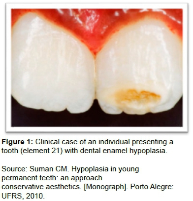
The first clinical case presented a brownish-colored cavity in the left upper central incisor, with a complaint of sensitivity and complaint of aesthetic damage, identified as hypoplasia (Figure 1).
1.What is your diagnosis for this tooth?
( ) Higid tooth
( ) Fluorosis
( ) Amelogenesis Imperfecta
( ) Enamel hypoplasia
( ) Caries Lesion
2.What treatment would you choose for this tooth?
( ) No need for treatment
( ) Preventive treatment (application of fluoride)
( ) Prophylaxis with pumice stone
( ) Prosthetic restorative treatment
-
( ) Facets
-
( ) Contact lens
( ) Restorative treatment. Which restorative materials would you use?
-
( ) Composite resin
-
( ) Photopolymerizable glass ionomer
CLINICAL CASE 01:
A 10 years old female patient with black color presents a brownish-colored cavity in the upper left central incisor, with reports of sensitivity and aesthetic impairment. Carefully examine the image and mark only one answer that represents your diagnosis and treatment decision.
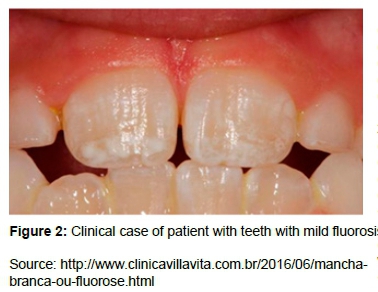
The second clinical case showed presence of white spots on the surface of the teeth, not requiring treatment, diagnosed as dental fluorosis (Figure 2).
The third clinical case showed defects, yellowish and stained teeth, sensitivity to touch, heat and cold in all teeth, diagnosed as amelogenesis imperfecta (Figure 3).
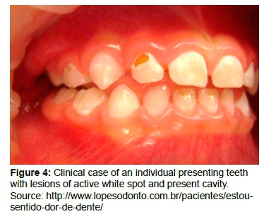
The fourth clinical case presented a child with decayed teeth (white patches and cavities) (Figure 4).
Inclusion criteria
The research included all dental academics from the fourth to the eighth levels enrolled in the disciplines of Dental Clinic, and the dentists professors of the undergraduate Dentistry course of Faculdade Meridional/ IMED, located in a city in the south of Brazil.
1. What is your diagnosis for this tooth?
( ) Higid tooth
( ) Fluorosis
( ) Amelogenesis Imperfecta
( ) Enamel hypoplasia
( ) Caries Lesion
2. What treatment would you choose for this tooth?
( ) No need for treatment
( ) Preventive treatment (application of fluoride)
( ) Prophylaxis with pumice stone
( ) Prosthetic restorative treatment
-
( ) Facets
-
( ) Contact lens
( ) Restorative treatment. Which restorative materials would you use?
-
( ) Composite resin
-
( ) Photopolymerizable glass ionomer
CLINICAL CASE 02:
A 22 years old female patient unhappy with the presence of white patches on the surface of her teeth. Carefully examine the image and mark only one answer that represents your diagnosis and treatment decision.
1. What is your diagnosis for this tooth?
( ) Higid tooth
( ) Fluorosis
( ) Amelogenesis Imperfecta
( ) Enamel hypoplasia
( ) Caries Lesion
2. What treatment would you choose for this tooth?
( ) No need for treatment
( ) Preventive treatment (application of fluoride)
( ) Prophylaxis with pumicet
( ) Prosthetic restorative treatment
-
( ) Facets
-
( ) Contact lens
( ) Restorative treatment. Which restorative materials would you use?
-
( ) Composite resin
-
( ) Photopolymerizable glass ionomer
CLINICAL CASE 03:
A female patient, 18 years old, had aesthetic defects, yellowish and stained teeth, sensitivity to touch, heat and cold in all teeth. Carefully examine the image and mark only one answer that represents your diagnosis and treatment decision.
1. What is your diagnosis for this tooth?
( ) Higid tooth
( ) Fluorosis
( ) Amelogenesis Imperfecta
( ) Enamel hypoplasia
( ) Caries Lesion
2. What treatment would you choose for this tooth?
( ) No need for treatment
( ) Preventive treatment (application of fluoride)
( ) Prophylaxis with pumicet
( ) Prosthetic restorative treatment
-
( ) Facets
-
( ) Contact lens
( ) Restorative treatment. Which restorative materials would you use?
-
( ) Composite resin
-
( ) Photopolymerizable glass ionomer
CLINICAL CASE 04:
Child with deciduous affected dentition. Carefully examine the image and mark only one answer that establishes your diagnosis and treatment decision.
RESULTS
All the questionnaires answers were typed in a Database built specifically for the present research. Subsequently, the data were exported to the statistical program SPSS 20.0 and submitted to descriptive statistical analysis, in order to verify the frequencies of the answers and to compare with the correct answers.
Table 1 shows the frequencies of the demographic variables of the students and professors, where the predominant gender among students 76 (77.6%) were women, and among professors 12 (52.2%) were women. The mean age of the students was 21.6 years (dp 3.6), with a minimum of 18 and a maximum of 37, and among the professors, the mean was 40.6 years (dp 9.1), with a minimum of 28 and a maximum of 58. Among the students, 70 (71.4%) considered that they had difficulty in the differential diagnosis in clinical practice, while the professors, 11 (47.8%) presented difficulties. Towards the individual knowledge regarding the differential diagnosis among the presented lesions, the 48 (49.0%) students reported having little knowledge, while the teachers, 12 (52.2%) said they had sufficient knowledge to diagnose, according to Table 1.
Table 2 shows the frequencies of knowledge variables on diagnosis and decision making to treat the lesions of the clinical cases presented. Of the 98 students, the predominant correct diagnosis was that of fluorosis, where 92 (93.9%) answered it right. While the predominant correct treatment was that of caries lesions, where 85 (86.7%) chose a direct restoration (Table 2).
Table 3 shows the frequencies of knowledge variables on diagnosis and treatment decision making of lesions from the clinical cases presented. Of the 23 teachers, the predominant correct diagnosis was for caries lesion, 23 (100%). While the predominant correct treatment was of direct restoration in the case of hypoplasia, where 22 (95.7%) opted for this treatment (Table 3).
DISCUSSION
The defects that affect the enamel's surface present themselves with very similar characteristics, implicating in a more complex diagnosis. Knowledge of dental enamel anomalies by the dental surgeon is essential to determine the differential diagnosis and to establish appropriate therapy.
Dental anomalies constitute deviations from normality caused by changes in the tooth's embryological development that can affect several aspects, such as quantity, size, shape, position in the arch, color and internal structure6. Dental enamel hypoplasia, demineralization and dental fluorosis result in lesions in the dental enamel characterized by local or generalized white patches, which detract from the aesthetic aspect due to the unnatural appearance of the dental enamel. Furthermore, in the specific case of active demineralization, require immediate interceptor treatment, and the differential diagnosis of these is essential to establish appropriate therapy9. Although some injuries are less common to occur, the professional should be prepared to deal with the situations and provide support, both clinical and emotional for the affected patients10.
Sampling choice for this study is due to the fact that the dental professional faces daily dental injuries, in academic life and later in professional practice, whose different defects affect the dental tissues with difficult differentiation. The distinct description of students and professors was due to different level of knowledge and experiences between them.
In order to diagnose the alterations affecting the dental tissues and to choose a suitable treatment in clinical practice, it is known that a clinical examination, prophylaxis of the teeth to obtain a clean and plaque-free surface, a proper drying of the teeth and good lighting is required. However, in the present study, this procedure could not be performed in the data collection, once the collection instrument used were photographs of clinical cases presented to the participants.
In the clinical situation presented in this study, in the first group, composed by dental students, with the clinical case number one of enamel hypoplasia, there was some difficulty in diagnosing the lesion, hence being the second diagnosis with fewer correct answers. One of the facts that explain this result may be because individuals with teeth presenting enamel hypoplasia are not commonly found in clinical practice. However, as there is color change in teeth with enamel hypoplasia, this fact may lead to different diagnoses11.
Thus, the diagnosis of enamel hypoplasia can be complicated and may be confused with many other enamel alterations, such as hypomineralization, hypomaturation and hypocalcification, where the treatment of enamel hypoplasia varies according to the severity of the change and also with age and behavior of the child, thus the indicated treatment may vary from topical applications of fluoride, to restorative, rehabilitative and aesthetic procedures12. In the group number two, represented by the professors of the Dentistry course, the case with the presence of hypoplasia was diagnosed with greater ease, being the second diagnosis with the highest number of correct answers, which was different from the group of students.
Regarding the treatment of this lesion, the best option is the direct restoration with composite resin, due to affect a young individual and this material is able to supply the restorative needs with excellent esthetics and function for this case. Direct restorative techniques provide a conservative, aesthetic and functional treatment in a single session, minimizing the amount of dental tissue to be removed in a tooth already compromised by enamel change13.
The treatment with direct adhesive restorations for the enamel hypoplasia presents as advantages, the low time of treatment, the ease of execution, satisfactory esthetics and low cost, because, using resinous dental materials it is possible to restore the dental anatomy and create a natural appearance of the teeth, restoring characteristics such as color, translucency, matrix, chroma and value11. Therefore the direct restoration was the treatment option of greater choice by students and professors of the present research.
A survey14 conducted to verify the knowledge and treatment decision of enamel defects by general practitioners and pediatric dentists has similar results to those obtained in the present study. In the two cases of enamel defects presented, most professionals did not identify hypoplasia and opacity. In the case of opacity, the diagnoses reported by most professionals were analogous to other structural defects of enamel, such as hypoplasia and fluorosis, and in the case of hypoplasia, many professionals believed it to be a decayed tooth14. Regarding the treatment, the results were equivalent with the answers of the group of students of the present study, where the majority opted for the direct restoration with composite resin.
Although fluoride is important for the control of dental caries, there is a risk of the occurrence of dental fluorosis where there is excessive intake of fluoride, dental fluorosis is dose-dependent and is related to the constant concentration of fluoride in the blood during dental formation. The greatest concern is the association to children's use of fluoridated water and fluoridated dentifrices, as well as the availability of fluoride in other sources, such as foods, teas and others15.The fluorosis, represented by clinical case number two, in the group of students was the diagnosis with the highest number of correct answers, this is due to the classic clinical characteristics of this lesion presented in the image, with well-defined white lines involving the homologous teeth. Dental fluorosis is more easily diagnosed because it occurs bilaterally and symmetrically, and its etiology is fluoride ingestion, which, associated with its clinical aspect, favors its diagnosis by anamnesis and a thorough examination of the patient1.
The results obtained in the diagnosis of fluorosis in the group of students were different from the results presented in the study done by Rigo et al.16, in the year 2015, in the same institution. Their research, composed of ten images of different degrees of severity of dental fluorosis, concluded that only three images were correctly diagnosed by the students, showing that an expressive number of students do not know how to diagnose it in clinical practice16. Thus, demonstrating that students are now acquiring more information for clinical decision-making. It can be inferred that currently, information is in many places beyond the academic context.
With the technological age and Internet use in academic life, it is easier to obtain qualified scientific data and information. Fontanella et al.17 describe that the use of information and communication technologies are tools of increasing importance for dentistry, as well as in other areas of health, promoting the use of new educational media that give students the ability to search for and select information, to learn independently and more autonomously and to solve problems.
In the group of teachers fluorosis was the lesion with the least number of correct answers. This fact may be associated with the presence of enamel opacities, occasionally confused with white caries lesion, however this lesion that precedes caries on smooth surfaces is usually easy to differentiate from opacities because it is associated with well demarcated biofilm deposits, adjacent to the gingival margin, extending along the lingual or palatine surfaces, on the contrary, the opacities do not have preferential place in the tooth and can be demarcated or diffuse14.
When dealing with fluorosis treatment, the interviewees presented greater difficulty, where only 40.8% of the students and 43.5% of the professors correctly answered that there was no need for treatment on the case. The question was that the patient had aesthetic complaint, but because it is a mild degree of fluorosis, there is no indication of treatment. The maximum to be done would be performing a microabrasion in the enamel, followed by dental whitening, only because the patient is unhappy with the spots. However, this treatment option was not contained in the questionnaire and the participants chose to treat the teeth affected by dental fluorosis. Although it is known that conservative therapeutic measures such as tooth whitening and enamel microabrasion may be beneficial in cases of mild fluorosis18. Invasive measures such as composite resin restorations, laminated veneers and total crowns are alternative treatments for cases of moderate to severe, aesthetically unpleasant and with loss of structure fluorosis. The therapeutic choice depends on the severity of dental fluorosis, that is, the clinical aspect. The opacities, when diagnosed, do not require restorative treatments, but should be chosen for their proservation or some of the conservative treatments (bleaching, microabrasion or macroabrasions), due to the low predisposition of this defect to dental caries, however, it cannot be neglected because some teeth with opacity may have their enamel ruptured, causing cavitation and allowing the adhesion of cariogenic bacteria14. However, there is a need for caution in the treatment of mild and moderate fluorosis, as the aesthetic impact caused by this condition is not directly proportional to its degree of severity.
Clinical case number three, represented by the amelogenesis imperfecta, was the most difficult lesion to diagnose for group one (students). Among several factors, this result is related to the little theoretical knowledge taught to the students, which have difficulty in diagnosing this type of lesion during clinical practice. While in group two, the vast majority correctly answered the diagnosis of amelogenesis imperfecta. According to study10, although amelogenesis imperfecta is a rare disease, where the formation of enamel is affected, the professional should be prepared to deal with the situation and provide both clinical and emotional support for these patients. Facing the treatment of this lesion, in both groups most of the participants opted for the correct treatment, referring to the indirect restoration.
However, even though the majority chose the treatment, it was noted that there was a resistance of the groups to perform an indirect restoration, which is due to the fact that today's dentists have a more conservative profile, in order not to subject the patient to unnecessary wear of teeth. Amelogenesis imperfecta is defined as a hereditary alteration of the enamel, which affects both dentitions, and also can cause tooth sensitivity, loss of vertical dimension and aesthetic compromise, thereby, several clinical situations that require resistance associated with aesthetics, and which previously were only solved with invasive prosthetic treatments, can now be solved with the latest restorative techniques and materials that allow less invasive restorative procedures19.
The diagnosis of dental caries and clinical decision making in dentistry are processes resulting from a balance of clinical and non-clinical factors related to the patient, procedure and dental surgeon20. Thus, the subjectivity of the professionals involved can interfere in these processes, even overlapping scientific knowledge based on evidence acquired in programs of Dentistry education.
The caries lesion, presented in clinical case number four, for group one was the second lesion with greater accuracy, this is due to the students' broad theoretical and practical knowledge regarding this condition and also the presented image shows a typical lesion of white stain on enamel with dentin cavitation. While in the group of teachers, this lesion obtained 100% accuracy, meaning that the professionals of the area have a great knowledge regarding these lesions. This is related to the fact that the carious lesion is a frequent disease in dental practice, where the professionals have extensive knowledge regarding their clinical characteristics and their development. Dental caries is one of the most common diseases in the world. It has a multifactorial nature, including essential factors (biofilm accumulation), determinant factors (exposure to sugars and fluorides) and modular factors (biological and social), concepts about the disease, incorporated during the professionals formation, can direct the type of conduct that will be adopted by them in caries control and treatment21.
Regarding caries treatment, many participants in both groups had doubts about the treatment of the lesion, since the patient was a child with caries lesions in enamel with advancement in dentin and whose treatment should be interventional, because the lesions were active and had cavities. The rehabilitation treatment for caries is a challenge for the Pediatric Dentistry because the child's age usually implies low cooperation during procedures, in addition, small amount of dental remnants, lower bond strength of the adhesive system to the deciduous tooth due to the histological and compositional characteristics of the tooth and difficulties inherent in the execution of the surgical technique and the restorative technique, hence making the rehabilitation treatment in children difficult and susceptible to failure22.
When the participants answered about the difficulties in the differential diagnosis in the clinical practice regarding the lesions affecting the dental tissues presented in the four clinical cases, in the sample of students, a large part reported having difficulty in the diagnosis decision (71.4%) and only 49 % reported having sufficient knowledge, while in the group of professors 47.8% reported having difficulties and 52.2% said they had sufficient knowledge to properly diagnose and treat the cases presented. Based on these results, it is believed that the students need a more theoretical and practical foundation against these lesions and should be approached more thoroughly by the professors during the undergraduate course, so that the students get a broad knowledge, eliminating the pertinent doubts. There is a great need to update the concepts of diagnosis and treatment of enamel defects among dental professionals14, which we corroborate with the results of the present study.
After the research, it was observed that the students found greater difficulty in two lesions (hypoplasia and amelogenesis imperfecta), while the teachers obtained smaller accuracy in the lesion of dental fluorosis. This fact may represent a need for greater knowledge in the diagnosis of structural defects that affect dental enamel. There is a great concern in educating capable professionals to recognize the alterations and to indicate the appropriate treatment16. Regarding the perception of lesions, the patient may not judge the defect as an aesthetic problem and mild fluorosis does not seem to be a concern. The dentist is advised to consider the patient's perception in order to avoid future disorders and treatments. However, when the treatment is proposed, the patient should also be aware of the limitations, especially in the more severe cases16.
One of the limitations of this study, that should be taken into account, is the fact that the answers were based on photographs of clinical cases, in a way that the figures used in the questionnaire were all focused only on the affected teeth, limiting the individual's visualization as an all, that is, without the interviewee obtaining a broader view of the situation addressed. Being just one image for each lesion, it may be a factor that has made it difficult to determine the diagnosis. A study reports that the positive aesthetic perception (acceptance) for the intraoral photographs is smaller than for the images that present the teeth in the context of the face (in a smile, for example), and is also associated with the distance of observation, concluding that intra-oral closes images may adversely affect aesthetic perception23. The photographs used in the present study were intra-oral images with enlarged dimensions, which may have contributed to a different view of oral reality.
As a contribution of this study, we could suggest the application of this methodology in several educational scenarios with the approach of other types of oral diseases, in order to verify the knowledge and the clinical decision making of qualified professionals or in habilitation for individuals care.
According to Tavares et al.24, knowledge is considered the starting point for decision making, in attempt to ensure the quality of the procedures performed. Dentistry courses must take up the challenge of modifying their curricula, since current teaching trends seek to integrate basic areas, propaedeutics and dentistry, and reduce the high burden of unnecessary technical knowledge and stimulate undergraduate students to strive for the correct diagnosis and the best treatment choice for their patients. It is important in academic curricula, from the first to the last year, that students can work these skills.
However, not forgetting that lifelong learning should be carried out individually by professors and health professionals, in order to sustain scientific knowledge and ensure the efficiency of transfering information to their students.
In conclusion: - Most of the students reported difficulty in the differential diagnosis when dealing with different lesions that affect the dental tissues and also, they lack sufficient knowledge to approach the clinical cases in the dental practice.
- The majority of the students were able to correctly diagnose the clinical case of an individual with fluorosis and the case with dental caries, but more difficulty in cases of amelogenesis imperfecta and dental enamel hypoplasia.
- In the group of teachers, all correctly diagnosed the case with caries lesions and most of them diagnosed the lesions of hypoplasia and amelogenesis imperfecta, whilst fluorosis caused greater difficulty of diagnosis.
- Both groups had difficulty in establishing treatment for fluorosis and amelogenesis imperfecta, while in the cases of enamel hypoplasia and caries lesion, most opted correctly for direct restoration.
REFERENCES
1. Sampaio FC, Forte FDS, Melo JM, Costa JDMC, Passos IA. Defeitos do esmalte: etiologia, características clínicas e diagnostico diferencial. Rev Inst Ciênc Saúde. 2007;25 (2):192-7. [ Links ]
2. Holffman RHS, Sousa MLR, Cypriano S. Prevalência de defeitos de esmalte e sua relação com cárie dentária nas dentições decídua e permanente. Cad Saúde Pública. 2007;23 (2):435-44. DOI: http://dx.doi.org/10.1590/S0102-311X2007000200020 [ Links ]
3. Nelson S, Albert JM, Lombardi G, Wishnek S, Asaad G, Kirchner HL, et al. Dental caries and enamel defects in very low birth weight adolescents. Caries Res. 2010;44(6):509-18. DOI: http://dx.doi.org/10.1159/000320160 [ Links ]
4. Bevilacqua FM, Sacramento T, Felício CM. Amelogênese imperfeita, Hipoplasia de esmalte e Fluorose dental-revisãode literattura. Rev Bras Multisdisc. 2010;13(2):136-48. DOI: https://doi.org/10.25061/2527-2675/ReBraM/2010.v13i2.146 [ Links ]
5. Ribas AO, Czlusniak GD. Anomalias do esmalte dental: etiologia, diagnostico e tratamento. Publ UEPG Cienc Biol Saúde. 2004;10(1):23-36. DOI: http://dx.doi.org/10.5212/publicatio%20uepg.v10i1.379 [ Links ]
6. Lanza MDS, Albuquerque NAR, Zica JSS, Rocha WMS, Ferreira RH, Lanza MD. Reabilitação funcionale estética de Amelogênese Imperfeita: relato de caso. Clinic Int J Braz Dent. 2016;12(2):164-71. [ Links ]
7. Fejerskov O, Nyvad B, Kidd EAM. Característica clínicas e histológicas da cárie dentária. In: Fejerskov O, Kidd EAM. Cárie dentária: a doença e seu tratamento clínico. São Paulo: Santos, 2005; p.71-97. [ Links ]
8. Instituto Brasileiro de Geografia e Estatística (IBGE). Pesquisa de orçamentos familiares. [cited 2015 Apr 15] Available from: https://www.ibge.gov.br/estatisticas-novoportal/sociais/saude/9050-pesquisa-de-orcamentos-familiares.html?=&t=o-que-e. [ Links ]
9. Pinheiro IVA, Medeiros MCS, Andrade AKM, Ruiz PA. Lesões brancas no esmalte dentário: como diferenciá-las e tratá-las. Rev Bras Patol Oral. 2003;2(1):11-18. [ Links ]
10. Azevedo MS, Goettems ML, Torriani DD, Romano AR, Demarco FF. Amelogênese imperfeita: aspectos clínicos e tratamento. Rev Gaúcha Odontol. 2013; 61(Supl.0):491-6. [ Links ]
11. Oliveira FV, Silva MFA, Nogueira RD, Geraldo-Martins VR. Hipoplasia de esmalte em paciente hebiátrico: relato de caso clinico. Rev Odontol Bras Cenral. 2015;24(68):31-6. [ Links ]
12. Campos PH, Santos VDRA, Guaré RO, Diniz MB. Dente hipoplásico de Turner: relato de casos clínicos. Rev Fac Odont Univ Passo Fundo. 2015;20(1):88-92. DOI: https://doi.org/10.5335/rfo.v20i1.4322 [ Links ]
13. Souza JB, Rodrigues PCF, Lopes LG, Guilherme AS, Freitas GC, Moreira FCL. Hipoplasia do esmalte: tratamento restaurador estético. Rev Odontol Bras Central. 2009;18(47):14-9. [ Links ]
14. Macedo-Costa MR, Passos IA, Oliveira AFB, Chaves AMB. Habilidade dos odontopediatras e clínicos gerais em diagnosticar e tartar defeitos do esmalte. Rev Gaúcha Odontol. 2010;58(3):339-43. [ Links ]
15. Marson FC, Sensi LG, Vieira LCC, Araújo FO. Clareamento dental associado à microabrasão do esmalte para remoção de manchas brancas o esmalte. Rev Dental Press Estét. 2007;4(1):89-96. [ Links ]
16. Rigo L, Lodi L, Garbin RR. Diagnóstico diferencial de fluorose dentária por discentes de odontologia. Einstein. 2015;13(4):547-54. DOI: http://dx.doi.org/10.1590/S1679-45082015AO3472 [ Links ]
17. Fontanella V, Schardosim M, Lara MC. Tecnologias de informação e comunicação no ensino da odontologia. Rev Abeno. 2007;7(1):76-81. [ Links ]
18. Oliveira BH, Milbourne P. Fluorose dentária em incisivos superiores permanentes em crianças de escola pública do Rio de Janeiro, RJ. Rev Saúde Pública. 2001; 35(3):276-82. DOI: http://dx.doi.org/10.1590/S0034-89102001000300010 [ Links ]
19. Silva W, Sousa LO, Montenegro G, Pinto T. A utilização de materiais adesivos no tratamento da amelogênese Imperfeita. Clinc Int J Braz Dent. 2012;8(2):178-86. [ Links ]
20. Mialhe FL, Silva RP, Ambrosano GMB, Pereira AC, Ferreira AC. Detecção e tratamento de lesões cariosas oclusais entre cirurgiões-dentistas do Sistema Único de Saúde. Rev Fac Odontol Univ Passo Fundo. 2007;12(3):29-34. [ Links ]
21. Ferreira-Nóbilo NP, Sousa MLR, Cury JA. Conceptualization of dental caries by undergraduate dental students from the first to the last year. Braz Dent J. 2014; 25(1):60-2. DOI: http://dx.doi.org/10.1590/0103-6440201302359 [ Links ]
22. Usha M, Deepak V, Venkat S, Gargi M. Treatment of severely multilated incisors: a challenge to the pedodontist. J Indian Soc Pedod Prev Den. 2007;25(Suppl):S34-6. [ Links ]
23. Baldani MH, Araújo PFF, Wambier DS, Strosky ML, Lopes CML. Percepção estética de fluorose dentária entre jovens universitários. Rev Bras Epidemiol. 2008; 11(4):597-607. DOI: http://dx.doi.org/10.1590/S1415-790X200800040000 [ Links ]
24. Tavares LFB, Bezerra IMP, Oliveira FR, Sousa LVA, Raimundo RD, Sousa EC, et al. Knowledge of Health Sciences undergraduate students in objective tests on Basic Life Support. J Hum Growth Dev. 2015;25(3):297-306. DOI: http://dx.doi.org/10.7322/jhgd.106002 [ Links ]
 Correspondence:
Izabel Barzotto
iza_barzotto@hotmail.com
Manuscript received: January 2018
Manuscript accepted: April 2018
Version of record online: June 2018


