SciELO - Scientific Electronic Library Online

 
vol.35 número1Cognitive, metabolic and cardiovascular effects of probiotics use among older adults: an umbrella review with meta-analysis and evidence mappingBack Health in Health Education: reflections on Musculoskeletal Fitness índice de autoresíndice de assuntospesquisa de artigos
Home Pagelista alfabética de periódicos  

Serviços Personalizados

Journal

artigo

Indicadores

Compartilhar


Journal of Human Growth and Development

versão impressa ISSN 0104-1282versão On-line ISSN 2175-3598

J. Hum. Growth Dev. vol.35 no.1 Santo André  2025  Epub 27-Jun-2025

https://doi.org/10.36311/jhgd.v35.17291 

ORIGINAL ARTICLE

Accelerated idioventricular rhythm in the adult: an update

Andrés Ricardo Pérez Riera, Wrote the paper, manuscript drafting, editing and submission, Design and coordination, All authors reviewed the results and approved the final version of the manuscripta 
http://orcid.org/0000-0003-4948-538X

Raimundo Barbosa Barros, Wrote the paper, manuscript drafting, editing and submission, All authors reviewed the results and approved the final version of the manuscriptb 
http://orcid.org/0000-0002-5113-3116

Daniel Henrique Kawatake de Souza, Wrote the paper, All authors reviewed the results and approved the final version of the manuscriptc 
http://orcid.org/0000-0003-0569-1181

Rodrigo Daminello Raimundo, Wrote the paper, manuscript drafting, editing and submission, All authors reviewed the results and approved the final version of the manuscriptd 
http://orcid.org/0000-0002-3043-0728

Samuel Sclarovsky, Wrote the paper, All authors reviewed the results and approved the final version of the manuscripte 

Kjell Nikus, Wrote the paper, manuscript drafting, editing and submission, Design and coordination, All authors reviewed the results and approved the final version of the manuscriptf 
http://orcid.org/0000-0002-9345-9851

aUniversidade Nove de Julho (Uninove), Campus Mauá, SP, Brazil; Hcor, Sao Paulo, Brazil. arperezriera@gmail.com

bCentro Coronariano, Hospital de Messejana Dr. Carlos Alberto Studart Gomes, Fortaleza, CE, Brazil. raimundobb@uol.com.br

cResidencia Médica em Clínica Médica Conjunto Hospitalar do MandaquiBrazil

dLaboratório de Delineamento de Pesquisa e escrita Científica Centro Universitário Faculdade de Medicina do ABC, Santo André, SP, Brazil. rodrigo.raimundo@fmabc.br

eEmeritus Professor at Tel Aviv University. Tel Aviv, Israel. samuel_s@netvision.net.il

fHeart Center, Tampere University Hospital,Finland

8Faculty of Medicine and Health Technology, Tampere University, Tampere, Finland. kjell.nikus@sydansairaala.fi


Abstract

Introduction

accelerated idioventricular rhythm (AIVR) is a typically regular ventricular rhythm with a heart rate between 50 and 110 (-120) beats per minute, absence of P waves or sinus P waves with atrioventricular dissociation, and a wide QRS interval. It is most commonly observed in the setting of acute myocardial infarction, reperfusion therapy, and structural heart diseases.

Objective

This article aims to review and update the current knowledge on accelerated idioventricular rhythm (AIVR) in adults, addressing its pathophysiology, underlying mechanisms, clinical presentation, differential diagnoses, and prognostic implications. Additionally, it seeks to synthesize the latest scientific evidence on the topic, discussing advances in clinical management and potential therapeutic approaches.

Methods

a comprehensive literature search was conducted using databases such as PubMed, Scopus, and Web of Science to identify recent studies on AIVR in adults. Articles discussing its mechanisms, clinical presentation, prognosis, and treatment approaches were analyzed and synthesized.

Results

AIVR is generally considered a benign arrhythmia, often self-limiting and not requiring specific treatment. However, it may indicate underlying cardiac pathology and, in some cases, contribute to hemodynamic instability. The differential diagnosis includes other ventricular arrhythmias, such as ventricular tachycardia, which necessitates careful electrocardiographic analysis. Current management strategies focus on treating the underlying condition rather than the arrhythmia itself.

Conclusion

AIVR in adults remains an important electrocardiographic finding, particularly in the context of acute coronary syndromes. While usually benign, recognition and appropriate differentiation from malignant arrhythmias are essential for optimal patient management. Further research is needed to clarify its prognostic implications and therapeutic considerations.

Key words: accelerated idioventricular rhythm; idioventricular rhythm; slow ventricular tachycardia

Authors summary

Why was this study done?

is usually a benign rhythm that doesn’t require direct antiarrhythmic therapy, while VT can be life-threatening.

What did the researchers do and find?

TImportantly, there is potential rate overlap between AIVR and some slow VT. AIVR should not be diagnosed solely based on ventricular rate; context is important. Other characteristics of AIVR are helpful for its correct diagnosis. Fusion and capture beats are much more frequent in AIVR than in VT, because the heart rate of AIVR is often close to that of the prevailing sinus rhythm. AIVR typically lacks the abrupt onset typical for VT.

What do these findings mean?

Accelerated idioventricular rhythm (AIVR) is currently defined as an enhanced ectopic ventricular rhythm with at least three consecutive ventricular beats, which is faster than intrinsic ventricular escape rhythm (≤40 bpm), but slower than VT (at least 100-120 bpm).

Key words: accelerated idioventricular rhythm; idioventricular rhythm; slow ventricular tachycardia

Highlights

Accelerated idioventricular rhythm (AIVR) is more commonly associated with reperfusion after myocardial injury.

AIVR is the most common reperfusion arrhythmia in humans.

AIVRs are distinguished from ventricular rhythms by its heart rates: less than 40 (ventricular escape) and faster than 120 (ventricular tachycardia).

AIVR is generally considered to be a benign abnormal heart rhythm. It is typically temporary and does not require treatment, though atrioventricular dyssynchrony can cause hemodynamic instability, which can be treated through overdrive pacing or atropine.

Key words: accelerated idioventricular rhythm; idioventricular rhythm; slow ventricular tachycardia

Resumo

Introdução

o ritmo idioventricular acelerado é um ritmo ventricular tipicamente regular, com uma frequência cardíaca entre 50 a 110 (-120) batimentos por minuto (bpm), ausência de ondas P ou ondas P sinusais com dissociação atrioventricular e um intervalo QRS largo. O mecanismo do ritmo idioventricular acelerado parece estar relacionado, na sua maioria, com o aumento do automatismo nas fibras His-Purkinje e/ou no miocárdio, por vezes acompanhado de excesso vagal e diminuição da atividade simpática. Quando o ritmo ventricular assume o controlo, é designado essencialmente por ritmo idioventricular.

Objetivo

revisar historicamente e conceitualmente o “ritmo idioventricular”, descrito pela primeira vez em 1911.

Método

foi efetuada uma revisão narrativa da literatura utilizando descritores relacionados com o ritmo idioventricular acelerado. As bases de dados electrónicas pesquisadas foram a LILACS e a MEDLINE.

Resultados

esta revisão abordou a história do ritmo idioventricular acelerado e fez uma revisão conceitual deste tipo de arritmia.

Conclusão

acredita-se que esses achados possam ser de grande valia para as equipes de saúde, contribuindo para melhores práticas clínicas em cardiologia.

Palavras-Chave: ritmo idioventricular acelerado; ritmo idioventricular; taquicardia ventricular lenta

INTRODUCTION

Other nomenclatures/synonymous terminology for accelerated idioventricular rhythm (AIVR): Idioventricular rhythm, “slow ventricular tachycardia (VT)”, “Reperfusion arrhythmia”, nonparoxysmal VT, accelerated isorhythmic ventricular rhythm, and the curious nomenclature “benevolent VT”1.

Definition: AIVR is a typically regular ventricular rhythm, with a heart rate between 50 to 110 (-120) beats per minute (bpm) absence of P waves or sinus P waves with atrioventricular dissociation, and a wide QRS interval. When the sinoatrial node is blocked or suppressed, latent pacemakers become active to conduct rhythm secondary to enhanced activity and generate escape beats that can be atrial, junctional or ventricular. When ventricular rhythm takes over, it is essentially called idioventricular rhythm2.

AIVR is currently defined as an enhanced ectopic ventricular rhythm with at least three consecutive ventricular beats, which is faster than normal intrinsic ventricular escape rhythm (≤40 bpm), but slower than VT (at least 100-120 bpm).

History: “Idioventricular rhythm” was first described by Sir Thomas Lewis in 1911 in his textbook “Mechanism of the Heart Beat” as the slow intrinsic ventricular rhythm not evident from the ECG during ordinary circumstances3. The topic was later expanded in his book entitled “The Mechanism and Graphic Registration of the Heart Beat. Whit Especial Reference to its Clinical Pathology published in 1920”. Lewis’s book is considered the first book on the electrocardiogram. The book is about the mechanism of the heartbeat and its clinical pathology. It includes (experimental and clinical) examples of graphic records and analysis, including AIVR, to help readers understand the subject.

Sir Thomas Lewis (1881-1945) was a British pioneer in cardiology.

1963: Phibbs, analyzed the diverse semantics and nomenclature such as interference and dissociations terms1.

1966: Marriot and Menendez introduced the current term for AIVR4 1969: Agustín Castellanos Jr. used the term “slow VT” to describe AIVR during an acute MI5.

1974. Rothfeld and Zucker described the polymorphic form of AIVR, which they called “Multiform accelerated idioventricular rhythm!!6.

AIVR and VT are both heart rhythms that originate in the ventricles, but they differ in heart rate and clinical implications:

Heart rate: AIVR has a heart rate between 50 and 110 (-120) beats per minute (bpm), while VT is faster than 100 bpm, normally >120 bpm.

Clinical implications: AIVR is usually a benign rhythm that doesn’t require direct antiarrhythmic therapy, while VT can be life-threatening.

ECG appearance: Both rhythms have wide QRS complexes on the ECG, but AIVR has a slower rate than VT.

Other differences between AIVR and VT include:

Hemodynamic stability: AIVR is mainly a hemodynamically stable rhythm that often occurs in acute myocardial infarction.

Complications: Rarely, AIVR can degenerate into VT or ventricular fibrillation (VF), resulting in hemodynamic instability and risk of sudden cardiac death.

AIVR is sometimes called a “slow ventricular tachycardia” because it has similar characteristics to VT, but a slower ventricular rate.

Importantly, there is potential rate overlap between AIVR and some slow VT. AIVR should not be diagnosed solely based on ventricular rate; context is important. Other characteristics of AIVR are helpful for its correct diagnosis. Fusion and capture beats are much more frequent in AIVR than in VT, because the heart rate of AIVR is often close to that of the prevailing sinus rhythm6,7. AIVR typically lacks the abrupt onset typical for VT.

AIVR is generally a transient rhythm, rarely causing hemodynamic instability and rarely requiring treatment. However, misdiagnosis of AIVR as slow ventricular tachycardia or complete heart block can lead to inappropriate therapies with potential complications. AIVR is often a clue to certain underlying conditions, like myocardial ischemia-reperfusion, digoxin toxicity, and cardiomyopathies8-11.

Pathophysiology

In most cases, the mechanism of AIVR appears to be related to enhanced automaticity in the His-Purkinje fibers and/or myocardium2, sometimes accompanied with vagal excess and decreased sympathetic activity12. Ischemia, reperfusion, hypoxia, drugs, and electrolyte abnormalities can all accelerate the phase 4 action potential depolarization rates in the His-Purkinje fibers and myocardium, leading to faster spontaneous cell depolarization (enhanced automaticity)8. When the enhanced automaticity in the His-Purkinje fibers or myocardium surpasses that of the sinus node, AIVR manifests as the dominant rhythm of the heart. Sinus bradycardia may facilitate the appearance of AIVR.

Under certain conditions such as acute ischemia and digoxin toxicity, triggered activity has been suggested as the mechanism for AIVR2.

Most AIVRs originate from a single focus. Occasionally, in patients with acute myocardial ischemia and myocarditis, AIVR can originate from multiple foci13-15.

Usually, AIVR is hemodynamically well tolerated due to its relatively slow ventricular rate. It is self-limited and resolves as sinus rate surpasses the rate of AIVR. Rarely, AIVR can degenerate into life-threatening ventricular arrhythmias. In patients with severe myocardial dysfunction, AIVR may lead to hemodynamic instability due to the loss of AV synchrony or the ventricular rate. Frequent AIVR may impair left ventricular function similar to the scenario with high rate of ventricular premature beats; left ventricular ejection fraction was negatively correlated with AIVR burden on Holter monitoring16.

Etiologies

AIVR can occur in apparently healthy individuals17-19. The most common cause of AIVR is myocardial reperfusion in acute myocardial infarction. The list of other etiologies is long, and includes the following:

  • Buerger disease20, or thromboangiitis obliterans (TAO), is a rare disease that causes inflammation and blockage of small and medium-sized blood vessels in the arms, legs, fingers, and toes. The exact cause of Buerger disease is unknown, but tobacco use is a central factor in its initiation and progression.

  • Congenital heart disease21 (CHD), including tetralogy of Fallot, ventricular septal defect, and Ebstein’s anomaly.

  • Dilated cardiomyopathy10 (DCM). Based on Holter findings, AIVR appeared to be a benign finding in patients with idiopathic DCM.

  • Myocarditis13. There are only a few case reports dealing with AIVR in myocarditis, and therefore, it is difficult to assess the clinical significance of this combination.

  • Drugs: digoxin toxicity9,22, cocaine toxicity23, and various anesthesia agents24-26.

  • Electrolyte abnormality such as hypo- and hyperkalemia (often associated with impaired renal function).

  • Post resuscitation27. In one study, post-resuscitation AIVR was associated with lower survival in successfully resuscitated out-of-hospital cardiac arrest victims.

  • AIVR in the acute myocardial infarction scenario.

Clinically, AIVR has been best studied in patients with acute ST-elevation myocardial infarction (STEMI)28,29. In the thrombolysis era, AIVR was noted to be a marker of reperfusion15,30. However, not all patients with reopened coronary artery have AIVR. In patients with acute myocardial infarction treated with primary percutaneous coronary intervention, the reported incidence of AIVR varied significantly, ranging from 15-42%, depending on methods of monitoring30.

Studies in patients with STEMI treated with primary percutaneous coronary intervention support that AIVR is a marker of reopening of an occluded coronary artery but is not necessarily a marker for complete reperfusion. In fact, AIVR seems to be associated with more extensive myocardial damage and delayed microvascular reperfusion30, although the mortality rates were similar in patients with and without AIVR. In primary percutaneous intervention patients, AIVR following reperfusion was associated with marked reduction in both systolic and diastolic blood pressure, irrespective of the infarct-related artery31.

In association with bundle branch block32. A case report described AIVR unmasking of the Brugada electrocardiographic pattern in a patient with right bundle branch block.

Epidemiology

  • Hingorani et al analyzed drug-free ambulatory ECG recordings from 1273 healthy volunteers (who had normal screening ECGs) from 22 phase 1 studies that were analyzed in a core ECG laboratory. AIVR was observed in 0.3% of these apparently healthy volunteers, while other types of arrhythmias were observed in a higher percentage33.

  • No racial preponderance exists, and men and women are equally affected.

  • No age predilection exists.

Prognosis

AIVR is a mostly self-limiting rhythm and typically has a benign prognosis, when AIVR rather than a slow VT is the true underlying entity2. The prognosis of patients with AIVR largely depends on their underlying conditions.

Morbidity/mortality

In general, AIVR does not significantly affect morbidity or mortality, which reflects that of the underlying condition causing the AIVR. In a retrospective observation study, AIVR was found to be associated with a lower 7-day survival rate in post-resuscitation patients compared to patients without AIVR27.

Patient history

History is helpful for identifying the underlying etiology for accelerated idioventricular rhythm (AIVR). AIVR per se may be an accidental finding in the 12-lead ECG, or more often, on Holter monitoring. The presence of the following conditions supports a potential diagnosis of AIVR:

Most patients with AIVR have chest pain or shortness of breath, symptoms related to myocardial ischemia. The arrhythmia typically appears at the time of myocardial reperfusion with drugs or coronary artery interventions8.

Some patients with AIVR have chest discomfort, shortness of breath, peripheral edema, cyanosis, clubbing, symptoms related to cardiomyopathy, myocarditis, and congenital heart diseases.

Occasionally, patients with AIVR have a history of using digoxin, some anesthetic agents, or cocaine.

Rarely, AIVR can occur in people without apparent heart disease and no identifiable triggers.

Physical Examination

There are no specific physical findings for accelerated idioventricular rhythm (AIVR). The following physical signs may be present:

  • Slow (< 55 bpm) or fast (>100 bpm) pulse rate.

  • Variable heart sound intensity and cannon A waves related to atrioventricular dissociation.

  • Some irregularity of heart rate/pulse rate due to competing sinus rhythm and AIVR.

  • Rarely, hypotension related to either AV asynchrony or relatively rapid ventricular heart rate during AIVR.

Diagnostic Considerations

Accelerated idioventricular rhythm (AIVR) is diagnosed based on its characteristic electrocardiographic findings. Its main differential diagnosis includes.

  1. Slow ventricular tachycardia, complete heart block,

  2. Junctional rhythm with aberrancy, supraventricular tachycardia with aberrancy, and

  3. Slow antidromic atrioventricular reentry tachycardia.

Example 1 : Electrocardiographic examples 

AIVR is a wide QRS ventricular rhythm with rate of 50-110 (120) bpm, often with variability during the episodes. However, AIVR should not be diagnosed solely by its ventricular rate because of the rate overlap between AIVR and some slow ventricular tachycardias. See the image below.

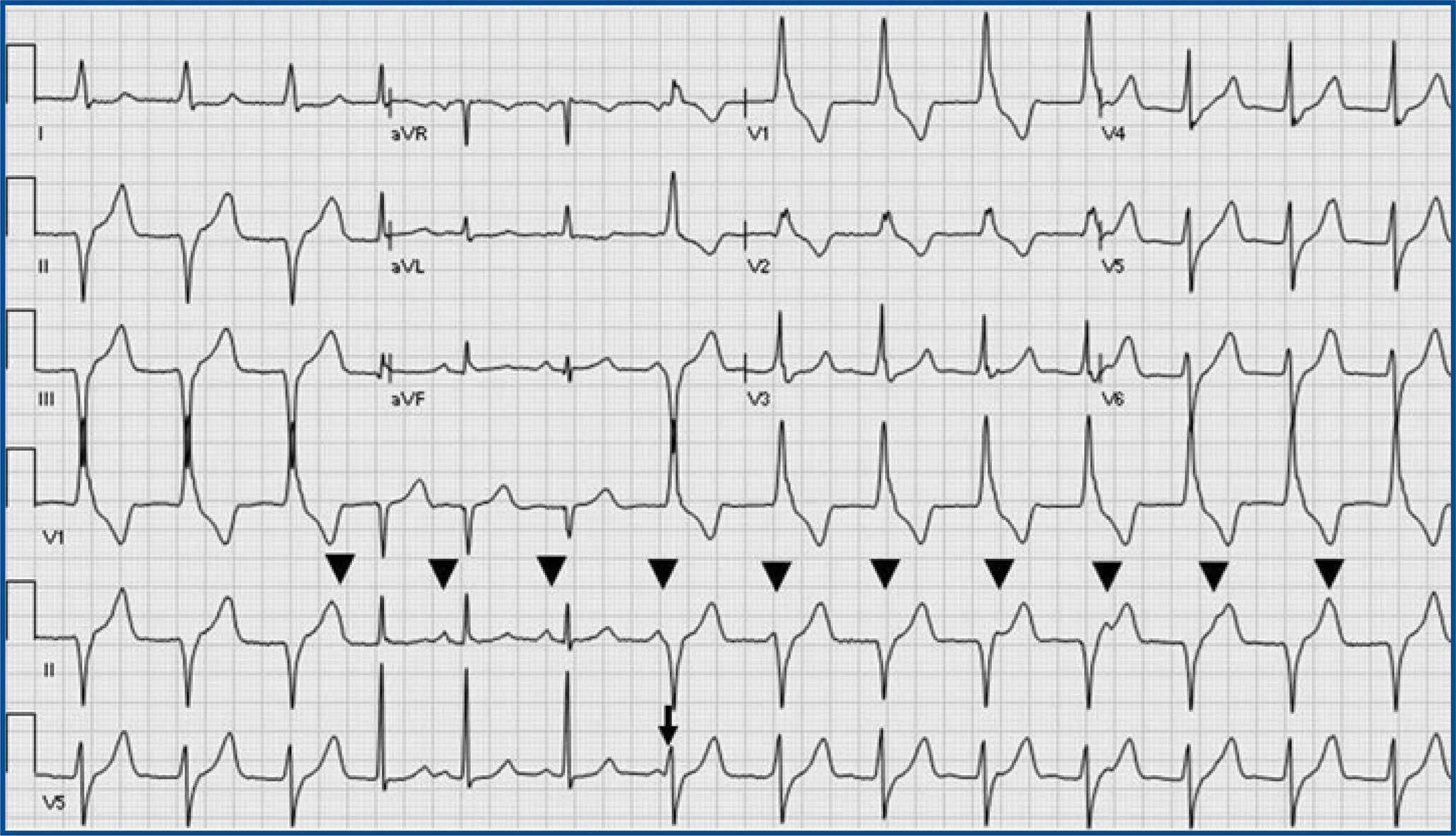
Accelerated Idioventricular Rhythm (AIVR) and sinus rhythm: AIVR starts and terminates gradually, competing with sinus rhythm. A possible ventricular fusion beat (arrow) and isoarrhythmic AV dissociation (arrowheads: sinus P waves) are present. During AIVR, ectopic ventricular rate is just faster than sinus rate. AIVR has a wide QRS morphology different from the QRS morphology in sinus rhythm.

Example 2  

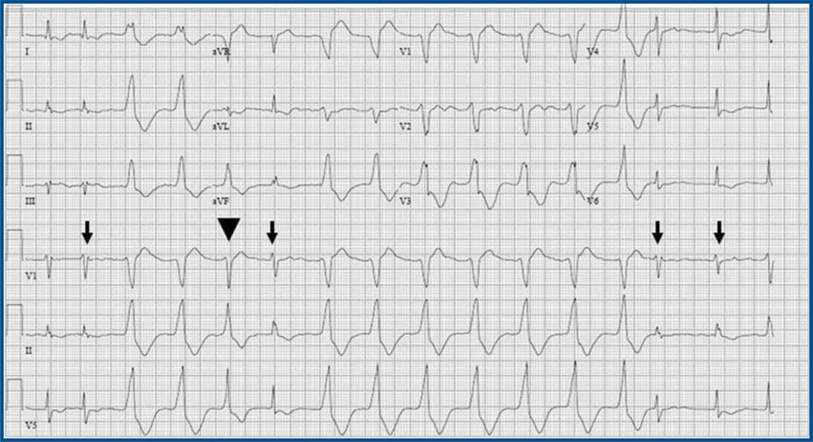
Accelerated Idioventricular Rhythm. AIVR in atrial fibrillation: AIVR starts and terminates gradually, competing with the ventricular capture beats (arrow) from atrial fibrillation. Ventricular fusion beat (arrowhead) is present. AIVR has a wide QRS morphology different from the QRS morphology of ventricular capture beats.

Example 3  

Junctional escape rhythm. Complete heart block with junctional escape rhythm: The AV dissociation in complete heart block is not isoarrhythmic AV dissociation, because the atrial rate is much faster than the ventricular rate of the junctional escape rhythm.

Example 4  

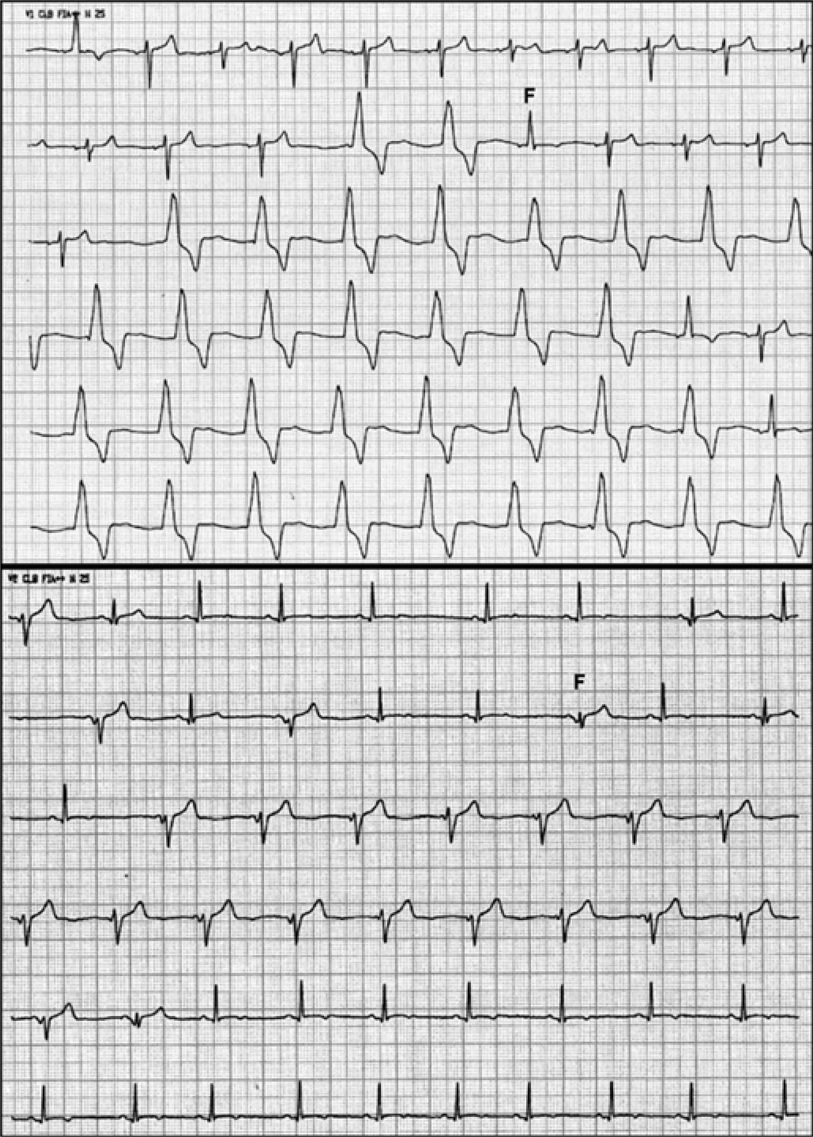
These ECGs belongs to a young, asymptomatic, Caucasian patient, without cardiovascular history, who practiced physical activities regularly. He had been treated with azithromycin for an upper airway infection. Both panels show sinus rhythm alternating with AIVR at approximately 60 bpm. Note fusion beats (F).

Example 5  

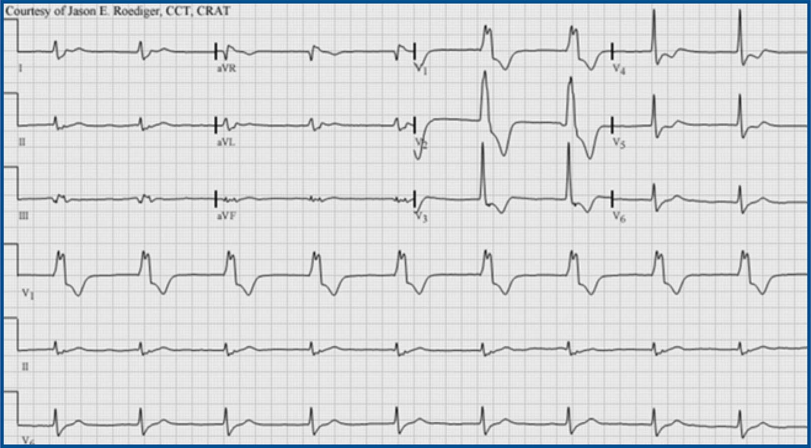
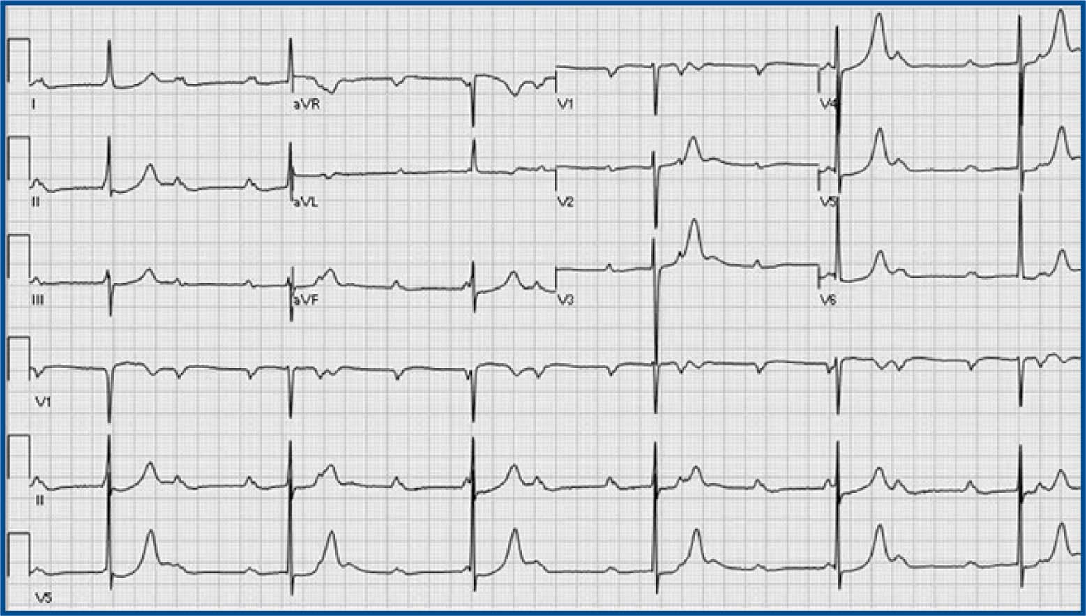
Accelerated idioventricular rhythm (AIVR), HR 55bpm, focus originating from the left ventricle: RBBB-like pattern in V1. Note the typical QRS morphology in lead V1 characteristic of ventricular ectopy from the LV. Monophasic R-wave with smooth upstroke and notching on the downstroke (i.e., the so-called taller left peak or “rabbit-ear”.) Contributed Courtesy of Jason E. Roediger (CC BY-SA 3.0 https://creativecommons.org/licenses/by-sa/3.0/deed.en).

Example 6  

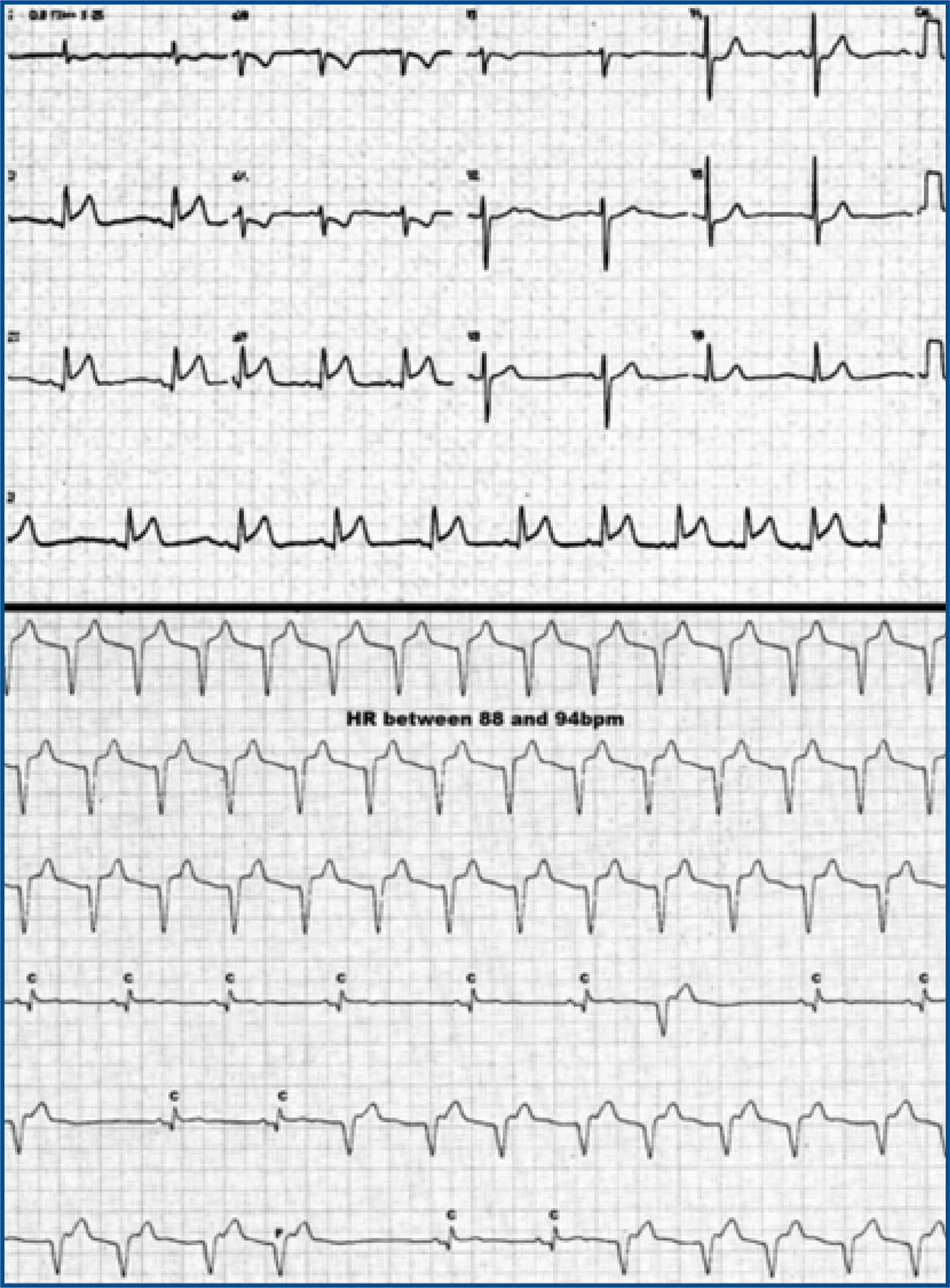
This ECG belongs to an elderly woman with acute ST-elevation myocardial infarction (superior panel). During thrombolytic administration, AIVR occurred (inferior panel). The heart rate is 88 bpm. AIVR alternates with sinus captures (C) and fusion beats (F). Sinus capture occurs when the sinoatrial node temporarily “captures” the ventricles, producing a QRS complex of normal duration. This can occur when the sinus rate is faster than the idioventricular rate. Fusion beats are hybrid complexes that occurs when a sinus and ventricular beat happen at the same time.

REFERENCES

1. Phibbs B. Interference, dissociation, and semantics. A plea for rational nomenclature. Am Heart J [Internet]. 1963 Feb;65(2):283-5. Available from: https://www.sciencedirect.com/science/article/pii/0002870363901650Links ]

2. Gangwani MK, Nagalli S. Idioventricular rhythm. In: StatPearls [Internet]. Treasure Island (FL): StatPearls Publishing; 2025. Available from: https://pubmed.ncbi.nlm.nih.gov/32119407/Links ]

3. Lewis T. The mechanism of the heart beat with especial reference to its clinical pathology [Internet]. Palala Press; 2015. 334 p. [ Links ]

4. Marriott HJ, Menendez MM. A-V dissociation revisited. Prog Cardiovasc Dis [Internet]. 1966 May;8(6):522-38. Available from: https://www.sciencedirect.com/science/article/abs/pii/S0033062066800142Links ]

5. Castellanos A Jr, Lemberg L, Arcebal AG. Mechanisms of slow ventricular tachycardias in acute myocardial infarction. Dis Chest [Internet]. 1969 Dec;56(6):470-6. Available from: https://www.sciencedirect.com/science/article/abs/pii/S0096021715346069Links ]

6. Rothfeld EL, Zucker IR. Multiform accelerated idioventricular rhythm. Angiology [Internet]. 1974 Jul;25(7):457-61. Available from: https://pubmed.ncbi.nlm.nih.gov/4843181/Links ]

7. Jakkoju A, Jakkoju R, Subramaniam PN, Glancy DL. Accelerated idioventricular rhythm. Proc (Bayl Univ Med Cent) [Internet]. 2018 Oct;31(4):506-7. Available from: https://pubmed.ncbi.nlm.nih.gov/31080367/Links ]

8. Goldberg S, Greenspon AJ, Urban PL, Muza B, Berger B, Walinsky P, et al. Reperfusion arrhythmia: a marker of restoration of antegrade flow during intracoronary thrombolysis for acute myocardial infarction. Am Heart J [Internet]. 1983 Jan;105(1):26-32. Available from: https://pubmed.ncbi.nlm.nih.gov/6849238/Links ]

9. Castellanos A, Azan L, Bierfield J, Myerburg RJ. Digitalis-induced accelerated idioventricular rhythms: revisited. Heart Lung [Internet]. 1975 Jan;4(1):104-10. Available from: https://pubmed.ncbi.nlm.nih.gov/1037686/Links ]

10. Grimm W, Hoffmann J, Menz V, Schmidt C, Müller HH, Maisch B. Significance of accelerated idioventricular rhythm in idiopathic dilated cardiomyopathy. Am J Cardiol [Internet]. 2000 Apr 1;85(7):899-904, A10. Available from: https://pubmed.ncbi.nlm.nih.gov/10758938/Links ]

11. Bonnemeier H, Ortak J, Wiegand UKH, Eberhardt F, Bode F, Schunkert H, et al. Accelerated idioventricular rhythm in the post-thrombolytic era: incidence, prognostic implications, and modulating mechanisms after direct percutaneous coronary intervention. Ann Noninvasive Electrocardiol [Internet]. 2005 Apr;10(2):179-87. Available from: https://pubmed.ncbi.nlm.nih.gov/15842430/Links ]

12. Grimm W. Accelerated idioventricular rhythm and bidirectional ventricular tachycardia. Cardiac electrophysiology: from cell to bedside [Internet]. 2004; Available from: https://cir.nii.ac.jp/crid/1571980075526907008Links ]

13. Nakagawa M, Hamaoka K, Okano S, Shiraishi I, Sawada T. Multiform accelerated idioventricular rhythm (AIVR) in a child with acute myocarditis. Clin Cardiol [Internet]. 1988 Dec;11(12):853-5. Available from: https://pubmed.ncbi.nlm.nih.gov/3233818/Links ]

14. Sclarovsky S, Strasberg B, Fuchs J, Lewin RF, Arditi A, Klainman E, et al. Multiform accelerated idioventricular rhythm in acute myocardial infarction: electrocardiographic characteristics and response to verapamil. Am J Cardiol [Internet]. 1983 Jul;52(1):43-7. Available from: https://pubmed.ncbi.nlm.nih.gov/6858925/Links ]

15. Hohnloser SH, Zabel M, Kasper W, Meinertz T, Just H. Assessment of coronary artery patency after thrombolytic therapy: accurate prediction utilizing the combined analysis of three noninvasive markers. J Am Coll Cardiol [Internet]. 1991 Jul;18(1):44-9. Available from: https://www.sciencedirect.com/science/article/pii/S0735109710802153Links ]

16. Wang L, Liu H, Zhu C, Gu K, Yang G, Chen H, et al. Clinical characteristics and therapeutic strategy of frequent accelerated idioventricular rhythm. BMC Cardiovasc Disord [Internet]. 2021 Sep 8;21(1):425. Available from: https://pubmed.ncbi.nlm.nih.gov/34496747/Links ]

17. Hiss RG, Averill KH, Lamb LE. Electrocardiographic findings in 67,375 asymptomatic subjects. III. Ventricular rhythms. Am J Cardiol [Internet]. 1960 Jul;6:96-107. Available from: https://pubmed.ncbi.nlm.nih.gov/13855920/Links ]

18. Chiale PA, Sicouri SJ, Elizari MV, Rosenbaum MB. Chronic idiopathic idioventricular tachycardia caused by slow response automaticity. Pacing Clin Electrophysiol [Internet]. 1987 Nov;10(6):1371-7. Available from: https://onlinelibrary.wiley.com/doi/abs/10.1111/j.1540-8159.1987.tb04972.xLinks ]

19. Riera ARP, Barros RB, de Sousa FD, Baranchuk A. Accelerated idioventricular rhythm: history and chronology of the main discoveries. Indian Pacing Electrophysiol J [Internet]. 2010 Jan 7;10(1):40-8. Available from: https://pubmed.ncbi.nlm.nih.gov/20084194/Links ]

20. Hsu PC, Lin TH, Su HM, Voon WC, Lai WT, Sheu SH. Frequent accelerated idioventricular rhythm in a young male of Buerger's disease with acute myocardial infarction. Int J Cardiol [Internet]. 2008 Jul 4;127(2):e64-6. Available from: https://pubmed.ncbi.nlm.nih.gov/17574693/Links ]

21. Nakagawa M, Yoshihara T, Matsumura A, Fusaoka T, Hamaoka K. Accelerated idioventricular rhythm in three newborn infants with congenital heart disease. Chest [Internet]. 1993 Jul;104(1):322-3. Available from: https://pubmed.ncbi.nlm.nih.gov/8325107/Links ]

22. Holzmann M, Reutter FW. Accelerated idioventricular rhythm with second degree v.a.-block and reentry (author's transl). Z Kardiol [Internet]. 1977 Jan;66(1):52-4. Available from: https://pubmed.ncbi.nlm.nih.gov/835325/Links ]

23. Jonsson S, O'Meara M, Young JB. Acute cocaine poisoning. Importance of treating seizures and acidosis. Am J Med [Internet]. 1983 Dec;75(6):1061-4. Available from: https://pubmed.ncbi.nlm.nih.gov/6316783/Links ]

24. Marret E, Pruszkowski O, Deleuze A, Bonnet F. Accelerated idioventricular rhythm associated with desflurane administration. Anesth Analg [Internet]. 2002 Aug;95(2):319-21, table of contents. Available from: https://pubmed.ncbi.nlm.nih.gov/12145043/Links ]

25. Chhabra A, Subramaniam R. Sudden appearance of idioventricular rhythm during inhalational induction with halothane in a child with congenital cataract. J Postgrad Med [Internet]. 2008 Oct;54(4):337-9. Available from: https://pubmed.ncbi.nlm.nih.gov/18953165/Links ]

26. Sato K, Miyamae Y, Kan M, Sato S, Yaegashi M, Sakanoue W, et al. Accelerated idioventricular rhythm following intraoral local anesthetic injection during general anesthesia. Anesth Prog [Internet]. 2021 Dec 1;68(4):230-4. Available from: https://pubmed.ncbi.nlm.nih.gov/34911065/Links ]

27. Tsai MS, Huang CH, Chen HR, Hsieh CC, Chang WT, Hsu CY, et al. Postresuscitation accelerated idioventricular rhythm: a potential prognostic factor for out-of-hospital cardiac arrest survivors. Intensive Care Med [Internet]. 2007 Sep;33(9):1628-32. Available from: https://pubmed.ncbi.nlm.nih.gov/17457573/Links ]

28. Hasin Y, Rogel S. Ventricular rhythms in acute myocardial infarction. Cardiology [Internet]. 1976;61(3):195-207. Available from: https://pubmed.ncbi.nlm.nih.gov/1009544/Links ]

29. Hoffman I, Zolnick MR, Bunn C. Transient post-reperfusion left bundle branch block and accelerated idioventricular rhythm with paradoxical QRS narrowing. J Electrocardiol [Internet]. 2014 Sep;47(5):705-7. Available from: https://www.sciencedirect.com/science/article/pii/S0022073614001575Links ]

30. Terkelsen CJ, Sørensen JT, Kaltoft AK, Nielsen SS, Thuesen L, Bøtker HE, et al. Prevalence and significance of accelerated idioventricular rhythm in patients with ST-elevation myocardial infarction treated with primary percutaneous coronary intervention. Am J Cardiol [Internet]. 2009 Dec 15;104(12):1641-6. Available from: https://pubmed.ncbi.nlm.nih.gov/19962468/Links ]

31. Delewi R, Remmelink M, Meuwissen M, van Royen N, Vis MM, Koch KT, et al. Acute haemodynamic effects of accelerated idioventricular rhythm in primary percutaneous coronary intervention. EuroIntervention [Internet]. 2011 Aug;7(4):467-71. Available from: https://pubmed.ncbi.nlm.nih.gov/21764665/Links ]

32. Elizari MV, Conde D, Baranchuk A, Chiale PA. Accelerated idioventricular rhythm unmasking the brugada electrocardiographic pattern: AIVR & Brugada ECG pattern. Ann Noninvasive Electrocardiol [Internet]. 2015 Jan;20(1):91-3. Available from: https://pubmed.ncbi.nlm.nih.gov/25040753/Links ]

33. Hingorani P, Karnad DR, Rohekar P, Kerkar V, Lokhandwala YY, Kothari S. Arrhythmias seen in baseline 24-hour Holter ECG recordings in healthy normal volunteers during phase 1 clinical trials: Arrhythmias in 24-hour Holter ECGs in healthy volunteers. J Clin Pharmacol [Internet]. 2016 Jul;56(7):885-93. Available from: https://pubmed.ncbi.nlm.nih.gov/26626443/Links ]

Funding: This article did not receive funding.

Received: November 2024; Accepted: December 2024; Published: April 2025

Corresponding author: arperezriera@gmail.com

Conflicts of Interest:

The authors declare no conflict of interest.

Creative Commons License this article is distributed under the terms of the Creative Commons Attribution 4.0 International License (https://creativecommons.org/licenses/by/4.0/), which permits unrestricted use, distribution, and reproduction in any medium, provided you give appropriate credit to the original author(s) and the source, provide a link to the Creative Commons license, and indicate if changes were made. The Creative Commons Public Domain Dedication waiver (https://creativecommons.org/publicdomain/zero/1.0/) applies to the data made available in this article, unless otherwise stated.